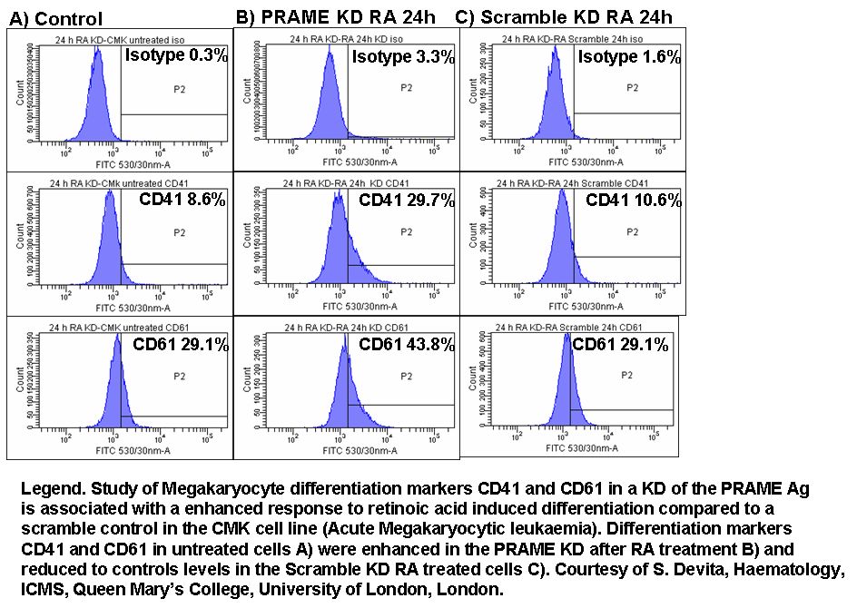
Immunophenotyping of CMK Megakaryocyte cell line
Differentiation markers, CD41 and CD61 were immunophentyped in response to retinoic acid treatment in a PRAME Ag Knock Down (KD) model and compared to a scramble control.
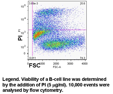
Cell viability can easily be determined in flow cytometry by adding one of DNA binding dyes at relatively low concentration to a population of cells. A common appproach is to use propidium iodide (PI) and 5 ug/ml; but there is a wide selection of dyes that can be used, which include 7-AAD, DAPI and To-Pro-3 all of these have to be used with caution as live cells will eventually take up these dyes if left to long, see figure and protocols.
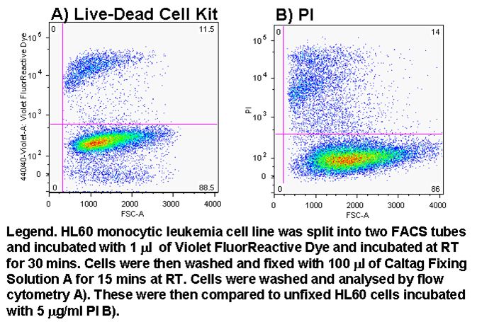
Determination of cell viability after fixing
The determination of cell viability when cells are infected with a pathogenic organism can be problematic given the requirement to fix the sample before flow cytometric analysis. However using the new Invitrogen reagent Violet Fluor-Reactive Dye allows the investigator to determine cell viabililty by flow cytometry by pre-incubation of cells with dye before fixing, see figure and protocol.